

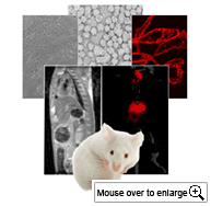
From molecules to whole animals Dr. Corbin’s Molecular imaging and Nanomedicine lab takes a multidisciplinary approach to studying some of the most challenging cancers. This collage of images highlights just a few of the many methods used in his research. These includes: an electron micrograph of targeted nanoparticles (top center) used for diagnostic and therapeutic applications; microscopy of cell culture systems (middle right and left) used to screen and evaluate novel drugs and imaging agents; finally in vivo whole animal imaging using high resolution MRI and high sensitivity near infrared fluorescent imaging(bottom right and left).
![]()
|
|||||||||||||

Ian R. Corbin, Ph.D., M.Sc.
Research Overview
Dr. Corbin’s molecular imaging and nanomedicine laboratory aims to develop innovative methodologies to enhance our understanding
and capabilities to detect and treat malignancies of the upper abdomen, namely of the liver and pancreas. His lab adopts a
multidisciplinary approach to research in which methods range from contrast agent synthesis and nanoparticle formulation, to cell
culture screening of targeted and therapeutic agents, to finally pre-clinical whole animal in vivo imaging and drug treatment trials.
Poised between basic research and clinical medicine Dr. Corbin’s research incorporates novel strategies and techniques from both
extremes in a reciprocal bench-to-bedside manner to both direct and advance his research endeavors.
Key areas of interest include:
- Molecular imaging methods for early detection of cancer;
- Development of nanoscale drug delivery systems for the treatment of hepatocellular carcinoma;
- Magnetic resonance spectroscopic evaluation of liver disease and cancer.
Contact: ian.corbin@utsouthwestern.edu
Research Interests
| Nanomedicine | Molecular Imaging |
| Magnetic Resonance Spectroscopy | Hepatocellular Carcinoma |
© The University of Texas at Dallas
