

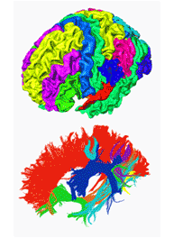
By integrating the information of cortical parcellation (upper image) from structural MRI and tractography from diffusion MRI (lower image), structural connectivity information can be obtained to study how the most complicated structure, the human brain, is “wired”.
![]()
|
|||||||||||||
Hao Huang, Ph.D., M.S.
Research Overview
Dr. Huang’s primary research interest is to use magnetic resonance imaging (MRI) and analysis techniques, especially those related to
diffusion imaging (including diffusion tensor imaging or DTI), to delineate the details of the brain anatomy. Dr. Huang’s lab is dedicated
to developing and applying these techniques to both answer biological questions and identify biomarkers for disease progression. For
clinical studies, physical parameters derived from DTI are used to quantitatively characterize structural abnormality of brains with
neurological or psychiatric diseases. Connectivity information from tractography of diffusion imaging and resting state functional MRI
is quantified to study how the human brain is “wired”. High resolution is pushed for MR microimaging at high magnetic field to obtain
both macro- and micro- structural information revealing the developmental pattern of human or animal brains.
Contact: hao.huang@utsouthwestern.edu
Research Interests
| Clinical application of DTI | Animal MRI |
| High b and HARDI diffusion MRI | Pulse program development |
| Brain development and connectivity with DTI | |