

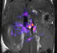
Functional imaging of the mouse pancreas during glucose stimulated insulin secretion (GSIS) by MRI. The colored overlay on this image of the mouse abdomen shows the tissue regions that become T1 enhanced by the Zn2+ ion sensor, GdDOTA-diBPEN, after a bolus injection of glucose to stimulate insulin secretion. Zn2+ is released from beta cells along with insulin and the free Zn2+ is detected by the MRI responsive sensor.
![]()
|
|||||||||||||||

A. Dean Sherry Ph.D.
Research Overview
My research program has two major thrusts. First, I have an interest in the impact of basic intermediary metabolism on human disease and in developing NMR and other physical methods to provide insights into cellular compartmentation, measurement of metabolic fluxes & flux control, and the influence of basic physiological parameters on metabolism in cells, tissues and whole organisms. Working with Craig Malloy, novel methods using the stable isotopes 13C and 2H as tracers and high resolution NMR techniques have been developed to allow measurement of multiple fluxes in vivo in animals and human subjects. 13C isotopomer NMR methods were initially developed in isolated, perfused hearts to measure substrate selection, anaplerosis, and TCA cycle flux, and to examine the influence of altered Ca2+ and redox state on metabolic fluxes. Methods were later developed to measure metabolic fluxes in live anesthetized animals and human subjects. With the addition of two human MR systems in 2006, our goal is to translate these methods to clinical studies of the influence of excess fatty acids on glucose and triglyceride storage and metabolism in skeletal muscle and liver in humans. 13C isotopomer methods have also been used to reveal that pyruvate cycling is an active metabolic process in the β-cell and that flux through this pathway correlates with glucose stimulated insulin secretion. Such studies may lead to new drugs to reverse the negative impact of accumulation of long chain fatty acids in β-cells on their ability of secrete insulin.
My second interest is the development of new molecular imaging agents that respond to physiology or metabolism and report that information by MRI. We have been involved with development of gadolinium complexes as non-specific extracellular MRI contrast agents since the early 1980’s and are now heavily involved with the next generation smart agents that respond to physiological parameters such as pH or for imaging the tissue distribution of the important metabolite, glucose. A new class of MR contrast agents was also recently discovered by our group that can be activated by specific RF pulses, called PARACEST agents. This new class of agents offers the potential to image a much broader spectrum of physiological/biochemical parameters including cell redox, enzyme activity, and tissue hypoxia.
Contact email: dean.sherry@utsouthwestern.edu
Research Interests
| MRS studies of intermediary metabolism in intact tissues |
PARACEST imaging agents |
| High field magnetic resonance spectroscopy of humans with metabolic diseases |
Molecular imaging of biological function |
| Gadolinium-based MRI contrast agents | Metabolic imaging agents |