

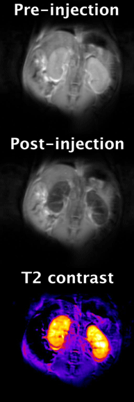
Magnetic resonance images of mouse kidneys at 9.4 Tesla showing the effects observed after injecting a 0.1 mmol/kg dose of a EuDOTA-based polymeric contrast agent. Uptake of the agent in the kidneys causes a 60% reduction in signal, making them appear dark (negative contrast). The negative contrast is caused by water molecule exchange between the Eu3+ ion and bulk water, which facilitates T2 exchange (T2exch). T2exch contrast is one on many novel methods used to further extend the functional and molecular imaging capabilities of MRI.
![]()
|
|||||||||

Todd C. Soesbe, Ph.D.
Research Overview
In vivo imaging of CEST (chemical exchange saturation transfer) and PARACEST (paramagnetic CEST) magnetic resonance contrast agents: CEST agents create contrast in MR
(magnetic resonance) images by exchanging their saturated Lanthanide bound protons with those of bulk water. Saturation is achieved by applying a 5 to 10 second long
frequency-specific pulse just before imaging. These agents have great potential to further extend the functional and molecular imaging capabilities of MR. Some
applications include measuring pH, angiogenesis, and glucose metabolism. We are currently investigating both monomer and polymeric Eu DOTA-(gly)4 based structures,
as well as a Eu DOTAM-based glucose sensor. We are also investigating several PARACEST imaging protocols including direct saturation and inversion-recovery (PCEST)
methods to help improve overall in vivo sensitivity.
In vivo imaging of liposomal Tm DOTMA: Liposomal delivery of MR contrast agents offers improved steady-state imaging and signal-to-noise due to their long blood circulation life-time. Also, the relative small size of the liposomes (≈ 100 nm in diameter) allows them to have direct uptake in certain tumor lines that exhibit "leaky" vasculature (e.g. MDA-MB-231 breast cancer cells). The 1H methyl group of Tm DOTMA has a chemical shift that is about -100 ppm away from water. This Tm DOTMA peak can be imaged using chemical shift selective (CHESS) techniques in which the water signal is completely absent. By using this method we can obtain "waterless" MR images where the only signal is due to the Tm DOTMA filled liposomes. This is analogous to images obtained in nuclear medicine where the signal is from the radioactive source only. Liposomal Tm DOTMA imaging has the potential to produce high resolution MR angiograms and tumor images that are not contaminated by the bulk water signal. Also, by labeling the outside surface of the liposome with a Gd-based pH sensor, the extracellular pH within a tumor can be mapped, which could lead to advances in therapeutic drug delivery
Contact email: todd.soesbe@utsouthwestern.edu
Research Interests
| In vivo imaging of CEST and PARACEST MR contrast agents | In vivo imaging of liposomal agents using MR |
| In vitro imaging of pancreatic beta cells using MR | MR probe design and construction |
| Preclinical SPECT detector design and construction | |