

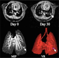
Typical respiratory- and cardiac-gated multi-slice gradient echo MR images (top) and maximum intensity projection (MIP) and volume rendering (VR) projection images (bottom) of a living mouse lung. The protocol was conducted at Day 0 and Day 30 in the same transgenic mouse to show the development of a sub-millimeter tumor (arrow), showing its reproducibility. Both MIP and VR images are generated from the 3D data set in which the image resolution (voxel size) is isotropic of (200 µm)3. One VR image (f) has sufficiently high transparency to exhibit the tumor.
![]()
|
|||||||||||||

Masaya Takahashi, Ph.D.
Research Overview
One of our major research focuses is on magnetic resonance imaging (MRI) of the lung. High-resolution computed tomography (HR-CT) is the standard for assessing lung morphology in pulmonary disorders, which incurs radiation exposure and yields data only about anatomy and not lung function. A report by the NIH/NHLBI identifies that an important challenge in pulmonary disorders is the development of more powerful, multi-variate methods for predicting regional outcome and individual responsiveness to particular therapies on the basis of clinical and laboratory characteristics.
MR imaging of the lung is extremely challenging due to multiple inherent difficulties associated with lung imaging. These challenges, include low proton density, severe magnetic field susceptibility, and respiratory and cardiac motion artifact have resulted in the delay in refining lung imaging as compared to MRI progress with other major organs. However, the recent development of fast MR imaging techniques along with more powerful hardware has made possible detailed, non-invasive imaging of pulmonary parenchyma, and pulmonary MRI is currently an active area of research. We have during the past years been dedicated to the development of new acquisition and processing methods by means of MR in animals and humans, permitting quantitative characterization of the pathophysiological change in pulmonary disorders, including lung cancer. We have developed a method to analyze mechanical function (motion/distortion) obtained from series of real-time MR images during normal and simulated breathing, and demonstrate in an animal model of emphysema that the new analysis technique accurately detects and maps the distributions of abnormalities with high spatial and temporal resolution. Another new MR method enables the imaging of lung parenchyma, which was unobservable previously using conventional MR methods. We are also developing methods to assess regional ventilation/perfusion imaging of the lung that is currently only available by scintigraphy, which is a new, complementary diagnostic dimension to functions and holds great promise. These projects should help establish a rigorous foundation for in vivo functional assessments of healthy lung and diseased lung, and can significantly broaden the scope of diagnostic capability without incurring the risks of radiation exposure associated with CT.
Contact email: masaya.takahashi@utsouthwestern.edu
Research Interests
| MRI of the lung | Molecular Imaging |
| Myelin Mapping |